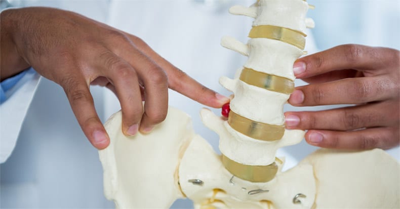
Low back pain (LBP) is VERY common condition, and research shows that up to 50% of the adult population in the United States will experience LBP in any three-month timeframe over the course of a year. Worse, low back pain can persist for months, years, and even longer, significantly reducing one’s ability to work, play, and enjoy life. So, let’s take a look at where LBP can come from…
ANATOMY: There are five lumbar vertebrae located just below the last rib and extending down to the sacrum. The FRONT of the vertebral column is made up of large box-shaped “vertebral bodies” that are strong and made to bear heavy weight. Between the vertebral bodies are shock-absorbing “intervertebral disks” that have a tough outer layer that surrounds a liquid-like center, giving it the ability to absorb vertical loaded pressure.
The spinal cord runs through the MIDDLE of the vertebra through the spinal canal. Nerves also exit the spine at each spinal level.
The BACK of the vertebra is made to protect the spinal cord. There are two gliding joints on the either side (called facet joints) of the vertebrae, which allow us to bend sideways, backwards, forward, or a combination of movements.
Below the lumbar spine sits the sacrum. The sacrum is wedged between the left and right wings of the pelvis, the ilia, forming the sacroiliac joint (SIJ). For many years, anatomists didn’t believe the SIJ could move and thus, could not be a pain generator. More recent research has concluded that not only is there movement in the SIJ but it may be the primary pain generator in up to 30% of lower back pain cases.
CASE STUDIES: Each of the above anatomical structures can be potential causes of LBP, and the presenting patient’s symptoms and clinical signs can help a doctor of chiropractic figure out what’s going on. For example, when a patient states, “My back kills me and the pain shoots down my leg when I bend over and feels better when I bend backwards and leg pain disappears,” this is most often caused by a herniated disk pinching a nerve in the low back.
In the above case, it’s important to examine the nerves that run down the leg, as the nerve can become damaged if too much pressure is exerted on the nerve for too long. Here, your doctor will ask you to walk on your toes and heels, check your reflexes at your knee and heel, and test your ability to feel sensations on the skin. If any of these tests reveal loss of function, the first goal of care will be to remove the pinch on the nerve to restore leg feeling and strength.
On the other hand, when a patient feels better bending over and worse bending backwards, the facet joints and/or the SIJ may be the culprit.


